
EDR 102
Positioning of the Teeth
- Posterior to focal trough
- If the patient’s anterior teeth are not positioned in the groove on the bite-block and are either too far back on the bite-block or posterior to the focal trough, the anterior teeth appear “fat” and out of focus on the radiograph.
- Anterior to focal trough
- If the patient’s anterior teeth are not positioned in the groove on the bite-block and are either too far forward or anterior to the focal trough, the teeth will appear “skinny” and out of focus.
What is the focal trough? (An imaginary curved space, shaped like a horseshoe.)
What happens if the jaws are outside this area? (The radiograph will not be sharp.) What happens if the jaws are inside this area? (The radiograph images will be well-defined.)
This patient is biting too far back on the bite stick.
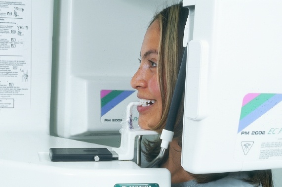
Be sure that the patient’s anterior teeth are in the grooves of the bite stick.
How do we obtain this film if a patient is missing all of his or her anterior teeth?
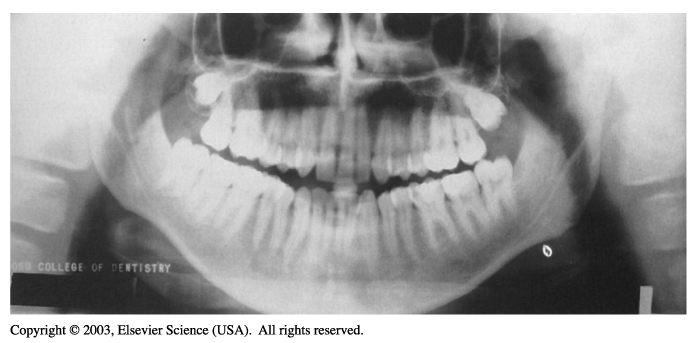
Anterior teeth appear widened and blurred on a panoramic radiograph when the patient is positioned too far back on the bite-block.
(From Haring J, Jansen L: Dental radiography: principles and techniques, ed 2, Philadelphia, 2000, Saunders.)

What should be done to correct this error? (Ask the patient to bite farther forward on the bite-block.)
Anterior teeth appear narrowed and blurred on a panoramic radiograph when the patient is positioned too far forward on the bite-block.
(From Haring J, Jansen L: Dental radiography: principles and techniques, ed 2, Philadelphia, 2000, Saunders.)
What should be done to correct this error? (Ask the patient to bite farther back on the bite-block.)
If the patient is not standing erect, superimposition of the cervical spine (arrows) may be seen on the center of the panoramic radiograph.
(From Haring J, Jansen L: Dental radiography: principles and techniques, ed 2, Philadelphia, 2000, Saunders.)

Always remind the patient to stand up straight and hold still during the exposure.
Positioning of the Spine
If the patient’s spine is not straight, the cervical spine will appear as a radiopaque artifact in the center of the film and obscure diagnostic information.
Extraoral Radiography
- Extraoral radiographs provide images of larger areas such as the skull and jaws.
- In some instances an extraoral film may be necessary in a handicapped patient who cannot open his or her mouth for film placement or in a patient with swelling or severe pain who is unable to tolerate the placement of intraoral films.
- Extraoral films are also useful in patients who are uncooperative and may refuse to open their mouths.
- Images seen on an extraoral films are not as clear or as well defined as the images seen on intraoral radiographs.
Extraoral radiography is not used to detect decay, periodontal disease, or periapical disease.
What are extraoral radiographs used for? (To detect fractures, lesions, diseases of the jaws, impacted teeth, and temporomandibular joint disorder.)
Equipment
- Extraoral radiographs may be taken with the use of a standard intraoral x-ray machine.
- Special head positioning and beam alignment devices can be added to the standard x-ray unit to aid patient positioning.
- Panoramic x-ray units may also be fitted with a special device known as a cephalostat.
- The cephalostat includes a film holder and head positioner that allow the operator to easily position the patient.
Where is the cephalostat inserted during patient positioning? (Just inside the ears.)
Will the cephalostat need to be disinfected?
Skull Radiography
- Lateral cephalometric projection
- Posteroanterior projection
- Water projection
- Submentovertex projection
- Reverse Towne projection
Interpreting these radiographs is difficult.
A lateral cephalometric view is the most common type of skull radiography.
Lateral cephalometric radiograph.
(From Haring J, Jansen L: Dental radiography: principles and techniques, ed 2, Philadelphia, 2000, Saunders.)

What is this radiograph used to evaluate? (Bones of the face and skull, including trauma, disease, or facial abnormalities.)
Used primarily for orthodontic purposes.
Posteroanterior skull radiograph.
(From Haring J, Jansen L: Dental radiography: principles and techniques, ed 2, Philadelphia, 2000, Saunders.)

What is this radiograph used to evaluate? (Facial growth and trauma, disease, and facial development.)
Submentovertex radiograph.
(From Haring J, Jansen L: Dental radiography: principles and techniques, ed 2, Philadelphia, 2000, Saunders.)

Not commonly used in dental offices.
What is this radiograph used to evaluate? (Position of the condyles, base of the skull, and fractures of the zygomatic arch.)
Reverse Towne radiograph.
(From Haring J, Jansen L: Dental radiography: principles and techniques, ed 2, Philadelphia, 2000, Saunders.)

Not commonly used in a dental office.
What is this radiograph used to evaluate? (Fractures of the condylar neck and ramus.)