
ODTP 101
Reasons a Patient Seeks Dental Care
- As a new patient
- For an emergency or a specific problem
- For consultation with a specialist
- As a returning patient for continued care
What does a “thorough examination” mean?
What is the clinical assistant’s role during a dental examination procedure? (To have the correct forms, instruments, and any additional supplies that may be needed by the dentist ready for use.)
Techniques Used For Assessment
- Visual evaluation provides an overall
assessment of existing conditions.- Soft tissue
- Tooth structure
- Restorations
- Missing teeth
- In palpation, the examiner uses the fingers and hands to evaluate hard and soft tissue.
- Instrumentation is the use of instruments to examine the teeth and surrounding tissues.
- Detection
- Probing
- Radiography, both intraoral and extraoral, provides indispensable tools for identifying:
- Decay
- Defective restorations
- Advanced periodontal conditions
- Pathologic conditions
- Developmental conditions
- Abnormalities
What does assessment mean? (Assessment is the process of making an official evaluation of a person or situation.)
How does the dentist use the explorer instrument? (With the use of a sharp pointed explorer, the dentist can detect imperfections in the enamel surface that may be the beginning of decay. The dentist will also use an explorer to evaluate existing restorations for their stability and integrity.)
(top) Example of an intraoral bite-wing radiograph.
(bottom) Example of an extraoral panoramic radiograph.


What are radiographs used for in a dental practice, and how does the dentist decide which radiographs to prescribe?
Radiographs have become indispensable tools in identifying decay, defective restorations, advanced periodontal conditions, pathology, developmental conditions, and other abnormalities.
- Intraoral imaging allows the use of a video system:
- To magnify an image for better evaluation
- For easier access to difficult areas
- For photocopying images for insurance purposes
- For case simulation or presentation
- For medical and legal documentation
What is intraoral imaging? (Intraoral imaging is similar to the use of a miniature video camera.)
This technique allows the dentist to use a computer monitor as a complement to a videocamera system, with a display of live video on a monitor screen.
An intraoral imaging system is used to make a diagnosis and educate the patient.

Notice that the members of the dental team are practicing infection control while using the intraoral camera.
- Intraoral and extraoral photography
- Provides a visual means of identifying and understanding specific problems.
In more comprehensive treatment, such as reconstructive or orthodontic procedures, before-and-after photographs will be taken to show the results.
Photographs are taken to provide a visual evaluation of the patient.

What is being used to pull back the lips and cheeks to allow the teeth to show?
Recording The Dental Examination
- Specific criteria that must be known before charting:
- Black classification of cavities
- Tooth diagrams
- Tooth-numbering systems
- Color coding
- Charting
Black Classification of Cavities

Developed by G.V. Black in the early 1900s, this standard classification system is universal to all dentists and is used to describe the location of decay and the best method for restoring the tooth. Black’s original classification included classes I through V. Class VI was added later.
What class in the Black system involves premolars and molars?
What class in Black’s system involves incisors?
Example of a geometric diagram for charting conditions of the mouth.
(From Gaylor LJ: The administrative dental assistant, ed 2, St Louis, 2007, Saunders.)
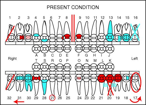
When referring to the diagram, remember that the teeth are presented from the perspective of looking into the patient’s mouth.
What dental charting systems are available? (A variety of diagram styles are available, but anatomic and geometric designs are most often used.)
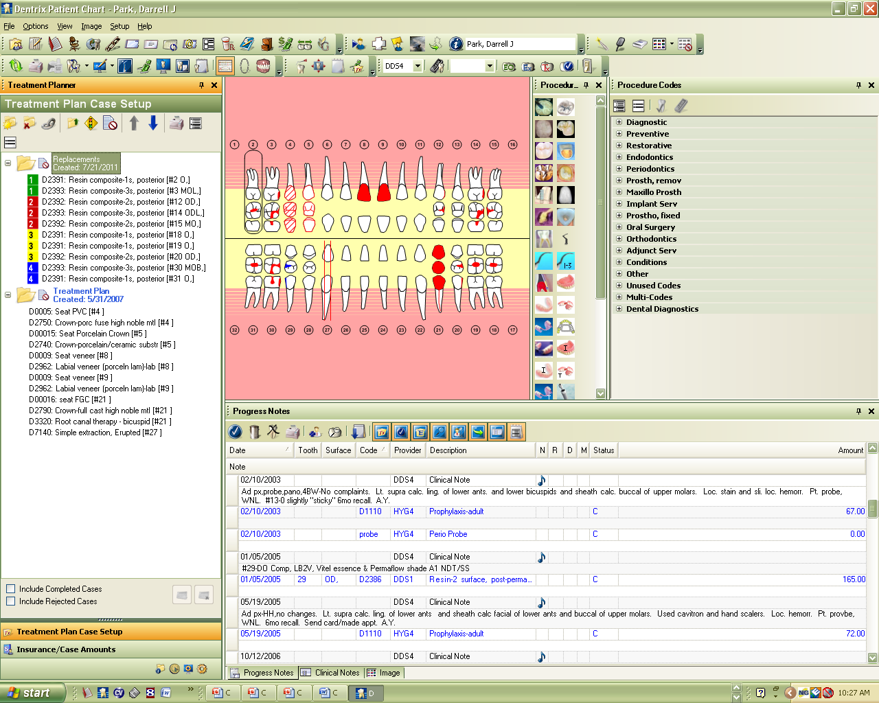
Universal Numbering System

How does this chart work? (This numbering system [1-32] begins with the maxillary right third molar and concludes at the mandibular right third molar.)
Is this an example of an anatomic or geometric chart?
Which charting system does your dentist use?
Which charting system do you think would work best for you or the patient?
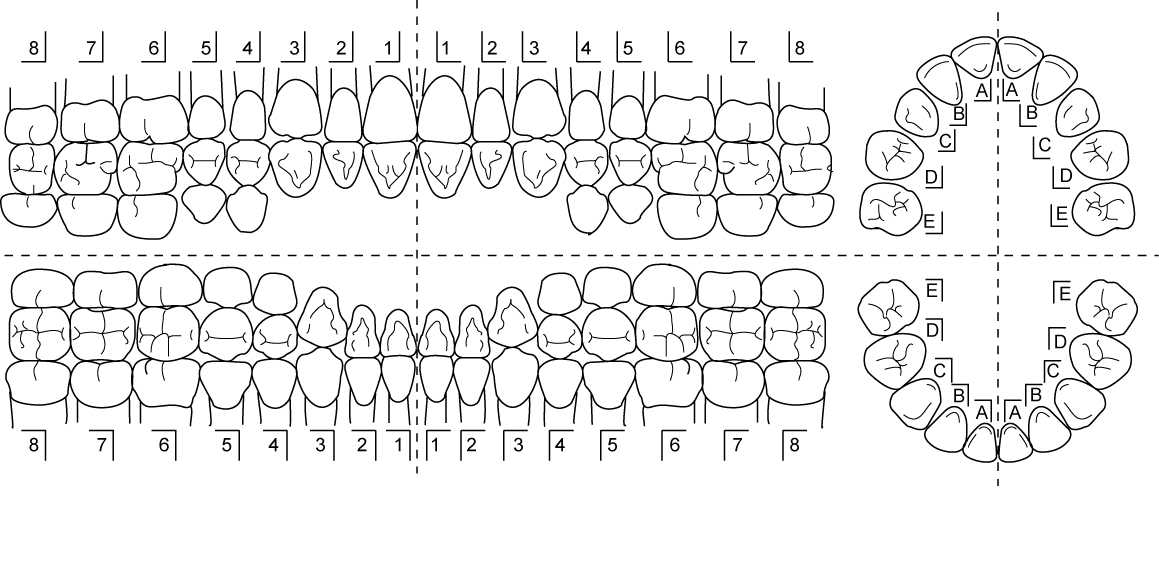
International Standards Organization System/Fédération Dentaire International System.

How does this chart work? (This numbering system assigns a two-digit number to each tooth; the first number is the quadrant and the second is the tooth.)
Is this an example of an anatomic or a geometric chart?
Fig. 28-9 C, Palmer notation system
How does this chart work? (This numbering system uses a bracket to designate the four quadrants of the mouth.)
Is this an example of an anatomic or a geometric chart?